Tuberculosis diagnosis
Tuberculosis is diagnosed by finding Mycobacterium tuberculosis bacteria in a clinical specimen taken from the patient. While other investigations may strongly suggest tuberculosis as the diagnosis, they cannot confirm it.

Diagnosis
A complete medical evaluation for tuberculosis (TB) must include a medical history, a physical examination, a chest X-ray and microbiological examination (of sputum or some other appropriate sample). It may also include a tuberculin skin test, other scans and X-rays, surgical biopsy.Medical history
The medical history includes obtaining the symptoms of pulmonary TB: productive, prolonged cough of three or more weeks, chest pain, and hemoptysis. Systemic symptoms include low grade remittent fever, chills, night sweats, appetite loss, weight loss, easy fatiguability, and production of sputum that starts out mucoid but changes to purulent[1]. Other parts of the medical history include prior TB exposure, infection or disease; past TB treatment; demographic risk factors for TB; and medical conditions that increase risk for TB disease such as HIV infection.Tuberculosis should be suspected when a persistent respiratory illness in an otherwise healthy individual does not respond to regular antibiotics.
Physical examination
A physical examination is done to assess the patient's general health and find other factors which may affect the TB treatment plan. It cannot be used to confirm or rule out TB.Microbiological studies

Sputum
Sputum smears and cultures should be done for acid-fast bacilli if the patient is producing sputum. The preferred method for this is fluorescence microscopy (auramine-rhodamine staining), which is more sensitive than conventional Ziehl-Neelsen staining. In cases where there is no spontaneous sputum production, a sample can be induced, usually by nebulized inhalation of a saline or saline with bronchodilator solution. A comparative study found that inducing three sputum samples is more sensitive than three gastric washings.Alternative sampling
In patients incapable of producing a sputum sample, common alternative sample sources for diagnosing pulmonary tuberculosis include gastric washings, laryngeal swab, bronchoscopy (with bronchoalveolar lavage, bronchial washings, and/or transbronchial biopsy), and fine needle aspiration (transtracheal or transbronchial). In some cases, a more invasive technique is necessary, including tissue biopsy during mediastinoscopy or thoracoscopy.PCR
Other mycobacteria are also acid-fast. If the smear is positive, PCR or gene probe tests can distinguish M. tuberculosis from other mycobacteria. Even if sputum smear is negative, tuberculosis must be considered and is only excluded after negative cultures.Other
Many types of cultures are available . Traditionally, cultures have used the Löwenstein-Jensen (LJ), Kirchner, or Middlebrook media (7H9, 7H10, and 7H11). A culture of the AFB can distinguish the various forms of mycobacteria, although results from this may take four to eight weeks for a conclusive answer. New automated systems that are faster include the MB/BacT, BACTEC 9000, and the Mycobacterial Growth Indicator Tube (MGIT). The Microscopic Observation Drug Susceptibility assay culture may be a faster and more accurate methodRadiography
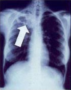
Chest X-ray
Tuberculosis creates cavities visible in x-rays like this one in the patient's right upper lobe.
In active pulmonary TB, infiltrates or consolidations and/or cavities are often seen in the upper lungs with or without mediastinal or hilar lymphadenopathy or pleural effusions ( tuberculous pleurisy). However, lesions may appear anywhere in the lungs. In disseminated TB a pattern of many tiny nodules throughout the lung fields is common - the so called miliary TB. In HIV and other immunosuppressed persons, any abnormality may indicate TB or the chest X-ray may even appear entirely normal.Abnormalities on chest radiographs may be suggestive of, but are never diagnostic of, TB. However, chest radiographs may be used to rule out the possibility of pulmonary TB in a person who has a positive reaction to the tuberculin skin test and no symptoms of disease.

Abreugraphy
A variant of the chest X-Ray, abreugraphy (from the name of its inventor, Dr. Manuel Dias de Abreu) was a small radiographic image, also called miniature mass radiography (MMR) or miniature chest radiograph. Though its resolution is limited (it doesn't allow the diagnosis of lung cancer, for example) it is sufficiently accurate for diagnosis of tuberculosis.Much less expensive than traditional X-Ray, MMR was quickly adopted and extensively utilized in some countries, in the 1950s. For example, in Brazil and in Japan, tuberculosis prevention laws went into effect, obligating ca. 60% of the population to undergo MMR screening.
The procedure went out of favor, as the incidence of tuberculosis dramatically decreased, but is still used in certain situations, such as the screening of prisoners and immigration applicants..
Tuberculin skin test (TST)
Two tests are available: the Mantoux and Heaf tests.Mantoux skin test
The Mantoux test for TB involves intradermally injecting PPD (Purified Protein Derivative) tuberculin and measuring the size of induration 48-72 hours later.
The Mantoux skin test is used in the 

Heaf test
For more details on this topic, see Heaf test.
The Heaf test was used in the The equivalent Mantoux test positive levels done with 10 TU (0.1 ml 100 TU/ml, 1:1000) are
- 0–4 mm induration (Heaf 0 to 1)
- 5–14 mm induration (Heaf 2)
- Greater than 15 mm induration (Heaf 3 to 5)
CDC classification of tuberculin reaction
An induration (palpable raised hardened area of skin) of more than 5–15 mm (depending upon the person's risk factors) to 10 Mantoux units is considered a positive result, indicating TB infection.- 5 mm or more is positive in
- HIV-positive person
- Recent contacts of TB case
- Persons with nodular or fibrotic changes on CXR consistent with old healed TB
- Patients with organ transplants and other immunosuppressed patients
- 10 mm or more is positive in
- Recent arrivals (less than 5 years) from high-prevalent countries
- Injection drug users
- Residents and employees of high-risk congregate settings (e.g., prisons, nursing homes, hospitals, homeless shelters, etc.)
- Mycobacteriology lab personnel
- Persons with clinical conditions that place them at high risk (e.g., diabetes, prolonged corticosteroid therapy, leukemia, end-stage renal disease, chronic malabsorption syndromes, low body weight, etc)
- Children less than 4 years of age, or children and adolescents exposed to adults in high-risk categories
- 15 mm or more is positive in
- Persons with no known risk factors for TB
- (Note: Targeted skin testing programs should only be conducted among high-risk groups)
BCG vaccine and tuberculin skin test
There is disagreement on the use of the Mantoux test on people who have been immunized with BCG. The US recommendation is that in administering and interpreting the Mantoux test, previous BCG vaccination should be ignored; the UK recommendation is that interferon-γ tests should be used to help interpret positive tuberculin tests, also, the UK do not recommend serial tuberculin skin testing in people who have had BCG (a key part of the US strategy). In their guidelines on the use of QuantiFERON Gold the US Centers for Disease Control and Prevention state that whereas Quantiferon Gold is not affected by BCG inoculation tuberculin tests can be affected. In general theUnder the
- Was in contact with another person with infectious TB
- Was born or has resided in a high TB prevalence country
- Is continually exposed to populations where TB prevalence is high.
Laboratory
Because of difficulties with the Tuberculin skin test, many laboratory methods of diagnosis are emerging. These tests have been reviewed in detailAdenosine deaminase
In 2007, a systematic review of adenosine deaminase by the NHS Health Technology Assessment Programme concluded "There is no evidence to support the use ofNucleic acid amplification tests (NAAT)
This is a heterogeneous group of tests that use the polymerase chain reaction (PCR) technique to detect mycobacterial nucleic acid. These test vary in which nucleic acid sequence they detect and vary in their accuracy. The two most common commercially available tests are the amplified mycobacterium tuberculosis direct test (MTD, Gen-Probe) and Amplicor (Roche Diagnostics). In 2007, a systematic review of NAAT by the NHS Health Technology Assessment Programme concluded that "NAAT test accuracy to be far superior when applied to respiratory samples as opposed to other specimens. Although the results were not statistically significant, the AMTD test appears to perform better than other currently available commercial tests." In a more recent before-after observational study, found that use of the MTD test reduce inappropriate tuberculosis therapy. The study found the accuracy of the MTD test as follows: Overall- sensitivity 92%
- specificity 99%
- sensitivity 99%
- specificity 98%
- sensitivity 62%
- specificity 99%
Full blood count
Although a full blood count is never diagnostic, normocytic anemia and lymphopenia are common. Neutrophilia is rarely found. [iron deficiency anemia may develop with isoniazid treatment] Urea and electrolytes are usually normal, although hypocalcemia and hyponatremia are possible in tuberculous meningoencephalitis due to SIADH. In advanced disease, hypoalbuminemia, hyperproteinemia, and hyperglobulinemia may be present.[9]Erythrocyte sedimentation rate is usually raised.
Interferon-γ release assays
Interferon-γ (interferon-gamma) release assays (IGRAs) are exciting new developments in TB infection testing. IGRAs are based on the ability of the Mycobacterium tuberculosis antigens for early secretory antigen target 6 (ESAT-6) and culture filtrate protein 10 (CFP-10) to stimulate host production of interferon-gamma. Because these antigens are not present in non-tuberculous mycobacteria or in any BCG vaccine variant, these tests can distinguish latent tuberculosis infection (LTBI).The blood tests QuantiFERON-TB Gold In Tube and T-SPOT.TB use these antigens to detect people with tuberculosis. Lymphocytes from the patient's blood are incubated with the antigens. These tests are called interferon γ tests and are not equivalent. If the patient has been exposed to tuberculosis before, T lymphocytes produce interferon γ in response. The QuantiFERON-TB Gold In Tube uses an ELISA format to detect the whole blood production of interferon γ with great sensitivity (89%). The distinction between the tests is that QuantiFERON-TB Gold quantifies the total amount of interferon γ when whole blood is exposed to the antigens(ESAT-6,CFP-10 and TB 7.7(p4)), whereas Guidelines for the use of the FDA approved QuantiFERON-TB Gold were released by the CDC in December 2005. In October 2007, the FDA gave approval of QuantiFERON-TB Gold In Tube for use in the
The enzyme-linked immunospot assay (ELISPOT) is another blood test available in the UK that may replace the skin test for diagnosis [11] [12] [13]. T-SPOT.TB, a type of ELISPOT assay,]counts the number of activated T lymphocytes that secrete interferon γ.
For diagnosing latent TB, three systematic reviews of IGRAs concluded the tests noted excellent specificity for the tests to distinguish latent TB from prior vaccination. According to a study from Korea, where there is a high prevalence of LTBI, QuantiFERON-TB Gold and T-SPOT.TB have good sensitivity but reduced specificity for diagnosing active TB, due to their ability to detect latent TB. In a recently published metaanalysis, with data from both developed and developing countries, QuantiFERON-TB Gold In Tube had a pooled sensitivity for active TB of 81% and specificity of 99.2%, whereas T-SPOT.TB had a pooled sensitivity of 87.5% and specificity of 86.3%. In head-to-head comparisons, the sensitivity of IGRAs surpassed TST. The authors concluded that IGRAs are "superior to the TST for detecting confirmed active TB disease
Public health
When someone is diagnosed with tuberculosis, all their close contacts should be screened for TB with a tuberculin skin test or a chest x-ray or both.Tuberculosis classification system used in the US
The current clinical classification system for TB (Class 0 to 5) is based on the pathogenesis of the disease.The U.S. Citizenship and Immigration Services has an additional TB classification (Class A, B1, or B2) for immigrants and refugees developed by the Centers for Disease Control and Prevention (CDC). The (Class) B notification program is an important screening strategy to identify new arrivals who have a high risk for TB.
Great post.This article is really very interesting and effective.I think its must be helpful for us. Thanks for sharing your informative lifescience journal.
ReplyDelete
ReplyDeleteI was diagnosed as HEPATITIS B carrier in 2013 with fibrosis of the
liver already present. I started on antiviral medications which
reduced the viral load initially. After a couple of years the virus
became resistant. I started on HEPATITIS B Herbal treatment from
ULTIMATE LIFE CLINIC (www.ultimatelifeclinic. com) in March, 2020. Their
treatment totally reversed the virus. I did another blood test after
the 6 months long treatment and tested negative to the virus. Amazing
treatment! This treatment is a breakthrough for all HBV carriers.